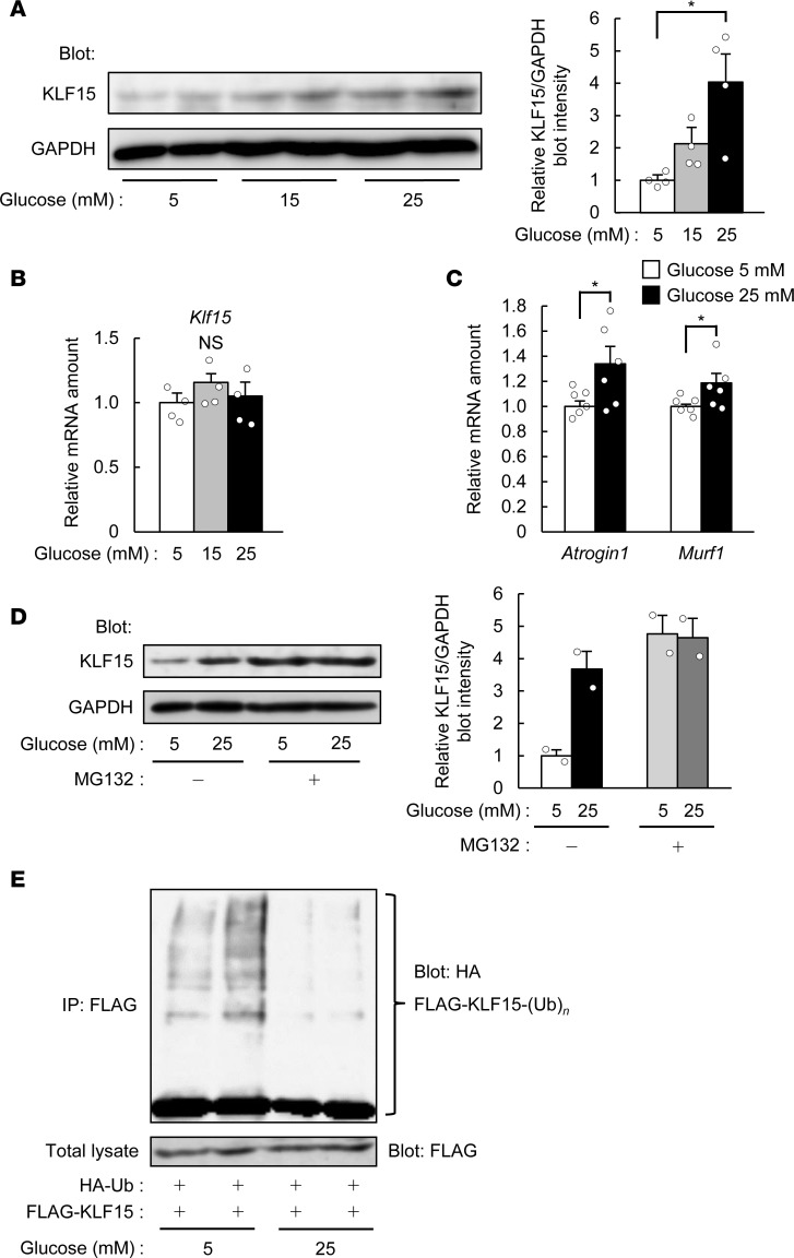
Figure 2. Glucose decreases the ubiquitination of, and increases the protein abundance of, KLF15.
(A and B) Immunoblot analysis of KLF15 protein (A) and quantitative RT-PCR analysis of Klf15 mRNA (B; n = 4) in C2C12 myotubes exposed to the indicated concentrations of glucose for 24 hours. In A, a representative blot and quantitative data (n = 4) are shown in the left and right panels, respectively. (C) Quantitative RT-PCR analysis of muscle atrophy–related gene expression for myotubes treated as in A. n = 6. (D) Immunoblot analysis of KLF15 in myotubes exposed to 5 or 25 mM glucose in the absence or presence of 15 μM MG132 for 6 hours. A representative blot and quantitative data (n = 2) are shown in the left and right panels, respectively. (E) C2C12 myoblasts expressing HA-ubiquitin (Ub) and FLAG-KLF15 were incubated with 5 or 25 mM glucose for 24 hours and then subjected to immunoprecipitation (IP) with antibodies against FLAG. The resulting precipitates were analyzed by immunoblot with antibodies against HA to detect polyubiquitinated [-(Ub)n] KLF15, and the original cell lysates were analyzed by immunoblot with antibodies against FLAG. Representative data from 3 independent experiments are shown. All quantitative data are means ± SEM for the indicated numbers of independent experiments. *P < 0.05; NS, not significant. Two-way ANOVA with Bonferroni’s post hoc test (A and B) or unpaired t test (C).

