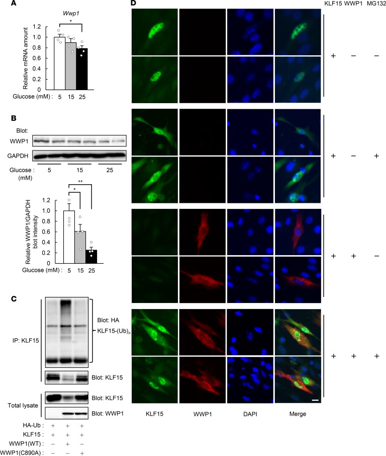
Figure 3. WWP1 regulates the polyubiquitination and abundance of KLF15.
(A and B) Quantitative RT-PCR analysis of Wwp1 mRNA (A; n = 4) and immunoblot analysis of WWP1 protein (B) in C2C12 myotubes exposed to the indicated concentrations of glucose for 24 hours. In B, a representative blot and quantitative data (n = 4) are shown in the left and right panels, respectively. (C) COS-7 cells transfected with vectors for HA-Ub, KLF15, and either WT or C890A mutant forms of WWP1 were subjected to immunoprecipitation with antibodies against KLF15. The resulting precipitates and the original cell lysates were analyzed by immunoblot as indicated. (D) Immunofluorescence analysis of KLF15 and WWP1 in C2C12 myoblasts transfected with vectors for these proteins and exposed to 15 μM MG132 for 6 hours as indicated. Nuclei were stained with DAPI. Scale bar: 10 μm. In C and D, representative data from at least 3 independent experiments are shown. All quantitative data are means ± SEM for the indicated numbers of independent experiments. *P < 0.05, **P < 0.01. Two-way ANOVA with Bonferroni’s post hoc test (A and B).

