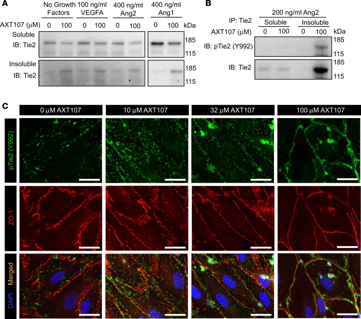
Figure 2. AXT107 alters Tie2 intracellular distribution.
(A) MEC lysates were treated with various growth factors and 100 μM AXT107 or DMSO vehicle and fractioned into Triton X-100–soluble and –insoluble pools. Blots were stained for total Tie2 (n = 3). (B) Representative images of Triton X-100–fractionated lysates immunoprecipitated for Tie2 and blotted for phospho-Tie2 (top) and total Tie2 (bottom); n = 3. (C) Immunofluorescence images of MEC monolayers treated with 200 ng/ml Ang2 for 15 minutes at varying concentrations of AXT107 and stained with DAPI (blue) and for phospho-Tie2 (Y992) (green) and ZO-1 (red) (n = 3). Scale bars: 25 μm.

