
Figure 1. Microglia Prune Developing CNS Synapses in a complement-dependent manner.
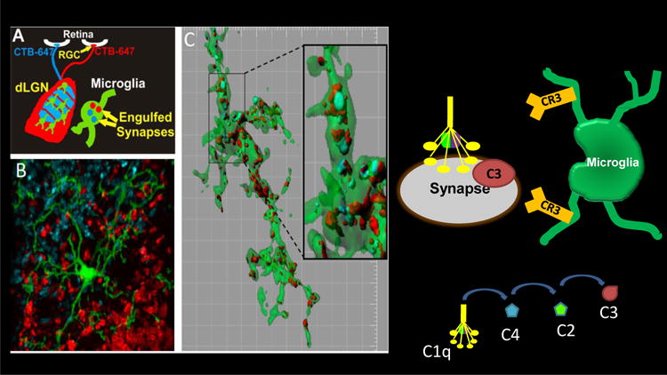
A Schematic of engulfment assay in which left and right eyes are injected with anterograde tracers, CTB-Alexa647 (blue) and CTB-Alexa488 (red), respectively B. Microglia (green) are visualized in the dLGN using a EGFP reporter mouse. C. Representative microglia (green) from the P5 dLGN surface rendered to visualize engulfed inputs (red and blue). [Grid line increments=5 μm] reveals presynaptic elements completely within microglia cytoplasm (green) (Schafer et al., Neuron 2012). Microglia engulfment of synapses is mediated, in part, by C3-CR3 phagocytic signaling as depicted on right.