. 2019 Apr 11;8:e45973. doi: 10.7554/eLife.45973

Figure 2. Activation of an exogenous reporter gene ZsGreen under the control of a HNF4α promoter by the CRISPR-assisted trans enhancer in multiple cells.

(a) Transcriptional activation of reporter gene ZsGreen in various cells transfected by different vectors. The florescence intensity of cells was analyzed by flow cytometry and is shown as the mean fluorescence intensity (MFI). Transfections: DVS, dCas9-VP64/sgRNA; DSC, dCas9/csgRNA-sCMV; DVSC, dCas9-VP64/csgRNA-sCMV. (b) Comparison between trans enhancer and VPR. Cells were transfected with three different transcriptional activation systems to activate reporter gene ZsGreen. The florescence intensity of cells was analyzed by flow cytometry and the number of cells with certain fluorescence intensity was counted. Transfections: Lipo, lipofectin; DVS, dCas9-VP64/sgRNA; DVPRS; dCas9-VPR/csgRNA; DVSC, dCas9-VP64/csgRNA-sCMV.

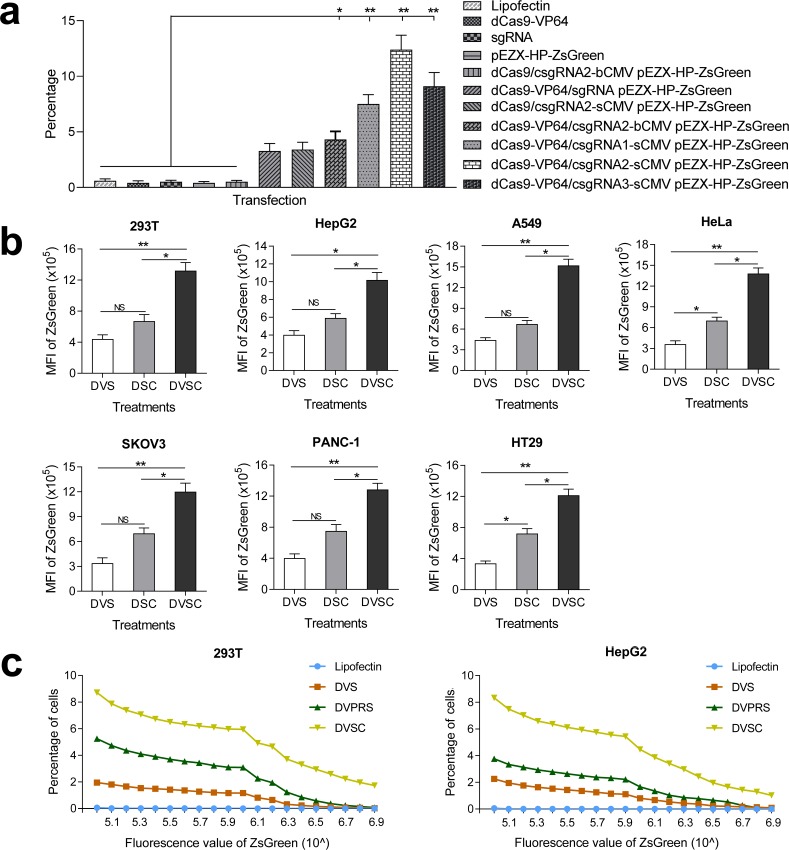
Figure 2.

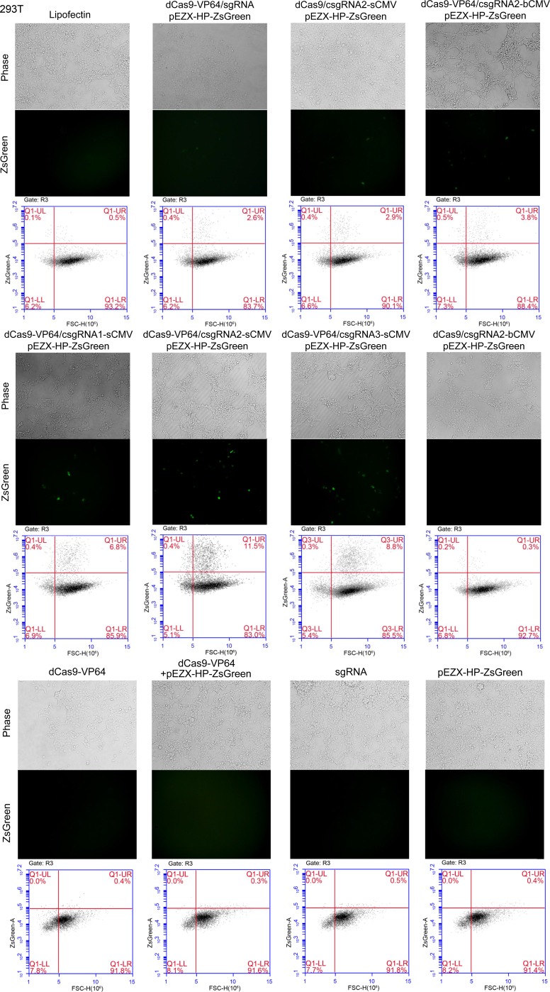
Figure 2—figure supplement 1. Activation of an exogenous reporter gene ZsGreen under the control of a HNF4α promoter by the CRISPR-assisted trans enhancer in 293 T cells.

Figure 2—figure supplement 1.

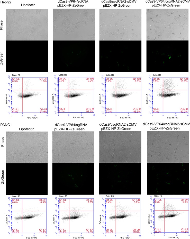
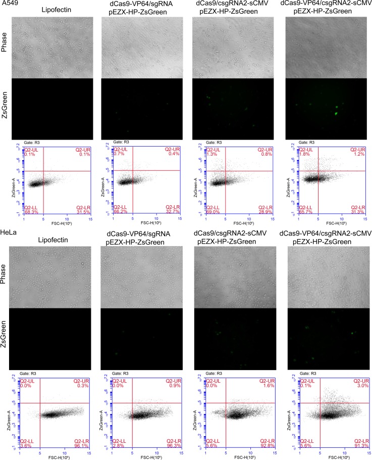
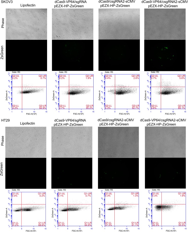
Cells were transfected by various vectors. Cells were photographed with a fluorescent microscope and their florescence was analyzed by flow cytometry. The reporter gene activation efficiency was indicated by the percentage of cells with green fluorescence over the threshold (cells in Q1-UR quadrant).
Figure 2—figure supplement 2. Activation of an exogenous reporter gene ZsGreen under the control of a HNF4α promoter by the CRISPR-assisted trans enhancer in HepG2 and PANC1 cells.

Figure 2—figure supplement 2.

Figure 2—figure supplement 3. Activation of an exogenous reporter gene ZsGreen under the control of a HNF4α promoter by the CRISPR-assisted trans enhancer in A549 and HeLa cells.

Figure 2—figure supplement 3.

Figure 2—figure supplement 4. Activation of an exogenous reporter gene ZsGreen under the control of a HNF4α promoter by the CRISPR-assisted trans enhancer in SKOV3 and HT29 cells.

Figure 2—figure supplement 4.