Figure 3:
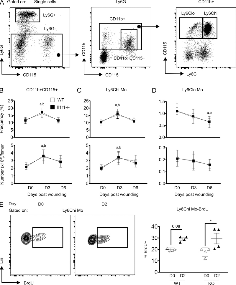
IL-1R1 deficiency does not alter skin wounding-induced monocyte expansion in BM. (A) Representative flow-cytograms showing gating strategy for flow-cytometry analysis of Mo subsets in BM. (B-D) Percentage (of total BMC; upper panels) and number (lower panels) of total Mo (Ly6G-CD11b+CD115+), Ly6Chi Mo (Ly6G-CD11b+CD115+Ly6Chi) and Ly6Clo Mo (Ly6G-CD11b+CD115+Ly6Clo) in BM. Results are represented as mean ± SD, n=7 mice for each strain and each time point. a, mean value significantly different from day 0 in WT; b, mean value significantly different from day 0 in Il-1r1−/−. (E) Representative flow-cytograms showing BrdU+ cells gated on Ly6Chi (left panel) and percentage of Ly6Chi BrdU+ cells (right panel). Results are represented as mean ± SD, n=3 to 4 mice for each strain and each time point. *p≤0.05.

