FIGURE 5:
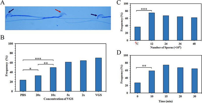
Sperm-VGS interaction in vitro and in vivo increases the frequency of acrosome-reacted (AR) sperm: in vitro response is dose-dependent. A) The acrosomal status of sperm was detected via Coomassie Blue staining after VGS co-incubation. Sperm with an intact acrosomal cap are shown by black arrows, while a missing cap after the acrosome reaction is shown by the red arrow. B) Sperm co-incubated in varying concentrations of the VGS (undiluted, 2x, 5x, 10x, 20x diluted VGS) or PBS for 30 min show a dose-response relationship. Using χ2 analysis, there is a significant (*=P<0.05, **=P<0.01, and ***=P<0.001) increase in AR between samples co-incubated with PBS and 20x / 10x dilution. C) Different numbers (12, 24, 36, 48 × 105) of sperm were deposited in the vaginas of superovulated mice and the rate of AR compared to the ex vivo control: †C (48 × 105). Chi-squared (χ2) analysis shows a significant (P<0.001) increase between the samples and control. D) Sperm (36 × 105) were deposited in the vaginas for different time periods (0–30 min). The percentages of AR sperm seen after 10 min and beyond were significantly (P<0.01) increased, compared to that of the start point (0 min). Increases between 10 and 30 min were insignificant.

