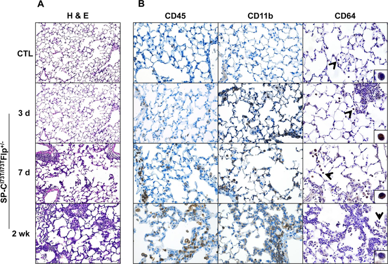
Figure 1. Parenchymal lung injury and alteration in immune cell composition induced by SP-CI73T expression.

(A) High power images of Hematoxylin & Eosin stained sections (100x) of control (tamoxifen treated SP-CWT/WTFlp+/+ or oil treated SP-CI73T/I73TFlp+/− mice) and SP-CI73T/I73TFlp+/− mice 3 days, 7 days and 2 weeks following intraperitoneal tamoxifen administration (250 mg/kg). (B) Immunohistochemical staining of lungs stained for CD45/immune cells (left panels), CD11b/migrating cells (center panels) and CD64/mature macrophages (right panels). Magnification: 200x; inset magnification 600x. Images shown are representative of 3–5 animals per group.
