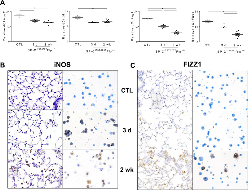
Figure 4. Phenotypic shift of BALF cells following SP-CI73T–induced injury.
(A) qPCR analysis of BALF cells collected from control (tamoxifen treated SP-CWT/WTFlp+/+ or oil treated SP-CI73T/I73TFlp+/− mice) and SP-CI73T/I73TFlp+/− mice 3 d, and 2 wk following intraperitoneal tamoxifen administration (250 mg/kg) for markers associated with M1 (Nos2 and Il6) and M2 (Arg1, Fizz1) activation. Data are represented as dCt relative to 18s and shown as mean ± SEM (N=3–6). *p< 0.05 compared to control SP-CWT/WTFlp+/+ or oil treated SP-CI73T/I73TFlp+/− mice SP-CI73T/I73TER−/− by One-Way ANOVA, followed by Tukey post-hoc test. (B-C) Immunohistochemical and cytospin staining of lungs for (B) iNOS and (C) FIZZ-1. Representative images are shown (200x; N=3–5) from control (tamoxifen treated SP-CWT/WTFlp+/+ or oil treated SP-CI73T/I73TFlp+/− mice) and SP-CI73T/I73TFlp+/− mice 3 d and 2 wk following intraperitoneal tamoxifen administration (250 mg/kg).

