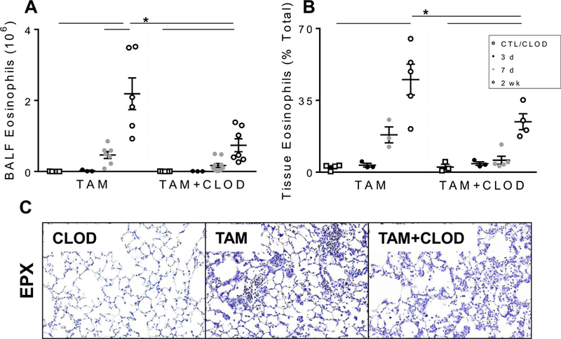
Figure 8. Peripheral monocyte depletion with intravenous clodronate liposomes results in altered eosinophil numbers in SP-CI73T mice.

(A-B) Changes in BAL and tissue SigFhiCD11b+CD11c− eosinophils from control (tamoxifen treated SP-CWT/WTFlp+/+ or oil treated SP-CI73T/I73TFlp+/− mice) and SP-CI73T/I73TFlp+/− mice 3 d, 7 d and 2 weeks following intraperitoneal tamoxifen (TAM, 250 mg/kg) and intravenous clodronate (CLOD, 150 µg/kg, 2 h post tamoxifen injection) administration. Data are represented as mean ± SEM (N=3–8). All analysis was considered significant *p< 0.05 compared to control SP-CWT/WTFlp+/+ or oil treated SP-CI73T/I73TFlp+/− mice SP-CI73T/I73TER−/− by One-Way ANOVA, using Tukey post-hoc test. C) Immunohistochemical analysis of vehicle and clodronate (150 µg/kg) treated control (tamoxifen treated SP-CWT/WTFlp+/+ or oil treated SP-CI73T/I73TFlp+/− mice) and SP-CI73T/I73TFlp+/− mice 2 weeks following intraperitoneal tamoxifen administration (250 mg/kg) lungs stained for eosinophil peroxidase (EPX). Representative 40x images from 3–5 separate animals at each condition are shown.
