Figure 2.

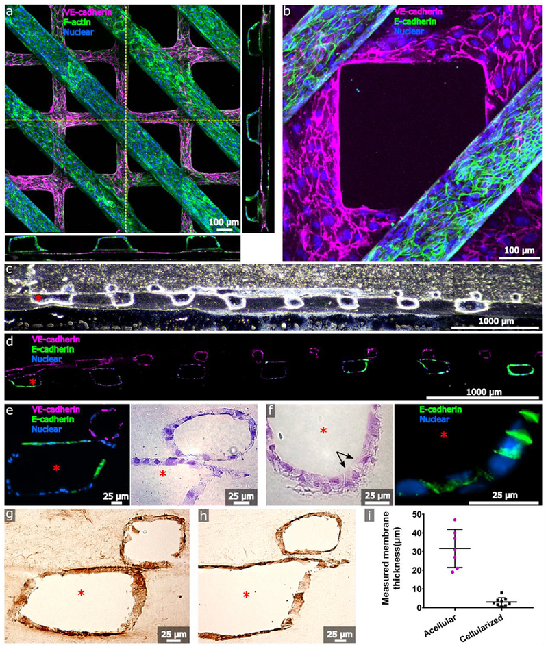
Cellularization of the human renal vascular-tubular unit (hRVTU). Top layers with grid geometry were seeded with P4-P6 human umbilical vein endothelial cells (HUVECs) and bottom layers with parallel-channel geometry were seeded with P1-P3 primary adult human renal cortical epithelial cells. A) Three-dimensional z-stack projection of an hRVTU with epithelial channel stained for F-actin and endothelial channel stained for VE-cadherin. Orthogonal images obtained along indicated planes (dashed yellow lines) are shown flanking the image and highlight the proximity of the two channels. B) 20× close-up of epithelial channel from A, stained for E-cadherin. C and D) Brightfield and fluorescent images of cryosectioned samples from an hRVTU following 14 days of culture and in situ immunostaining, at 10× magnification. E) Zoomed view of hRVTU from D (left), with H&E staining on a similar section shown adjacent (right). Channels show close apposition and heterogeneous junctional staining for E-cadherin. F) H&E staining of cryosectioned hRVTU reveals cuboidal cells with brush border-like structure (arrows) on the luminal face of the epithelium (left). Similar regions expressed basolateral E-cadherin (right). G) IHC staining of cryosectioned hRVTU for collagen IV. H) IHC staining of cryosectioned hRVTU for laminin. I) The membrane thicknesses of acellular devices were compared with devices following 14 days of cell culture. Solid lines indicate the mean and standard deviation for each condition. Red asterisk denotes the epithelial lumen, and representative colors for all staining are indicated within each panel.
