
Figure 4.
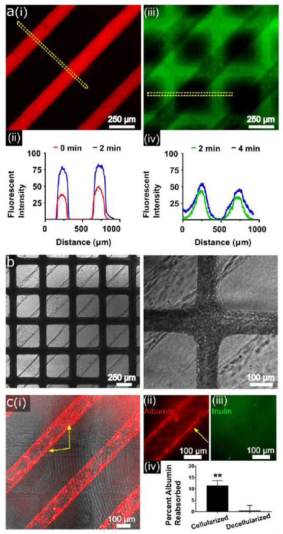
Functional analysis of human fetal and adult single-donor human renal vascular-tubular units (hRVTU). A) hRVTU created from human fetal renal cortical epithelial cells and kidney microvascular endothelial cells from the same donor sets were used for permeability assessment. Texas Red-labeled 70 kDa dextran was perfused into the epithelial channel during real-time imaging with 10 µm FITC-labeled 70 kDa dextran perfused into the endothelial 2 minutes later. (i) Epithelial channel is shown ~1 minute into perfusion and (ii) endothelial channel ~3 minutes into perfusion. (ii and iv) Fluorescent intensity over distance was calculated for the ROI shown in each image (dashed yellow lines) at two time-points using imageJ software (U.S. National Institute of Health, Bethesda, Maryland), demonstrating relatively little increase in width during the measured period. B) Blood from healthy volunteers was perfused into the endothelial channel of a hRVTU constructed as in A, under real-time imaging. No blood components can be seen within epithelial channels throughout ten minutes of blood perfusion. Left panel at 4× magnification, right panel at 10× magnification. C) hRVTU were created from adult renal cortical epithelial cells and human kidney microvascular endothelial cells from the same donor sets. After four days in culture device epithelial inlets were perfused with rhodamine-labeled human serum albumin and FITC-labeled inulin under real-time imaging. Following perfusion, devices were decellularized with trypsin, and perfusion repeated as an internal control. (i) Devices shown 15 minutes into perfusion show albumin (red) accumulating within cells. Yellow arrows highlight intracellular albumin accumulation within cells both on the top and side walls of vessels. (ii) Magnified image of a channel 30 minutes into perfusion shows albumin accumulating within cells, while inulin (iii) is not seen to accumulate intracellularly. Effluent was collected at one hour and run on a spectrophotometer, with albumin reabsorption calculated as noted in the main text. A two-tailed paired T test was done comparing albumin reabsorption in cellularized versus decellularized vessels (n = 3, bars indicate SEM and ** indicates P < 0.01).