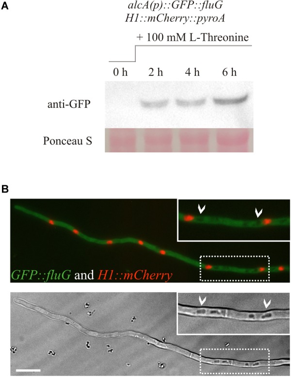
FIGURE 3.

(A) Western blot showing the protein expression of the N-terminally GFP-tagged FluG construct under the control of the alcA promoter. Induction of the alcA(p)::GFP::fluG strain shows an increment on the expression of GFP–FluG in time (2, 4, and 6 h). (B) Images obtained in the fluorescence microscopy show that FluG presents principally a cytoplasmic localization, excluded from the nuclei and is not detectable in vacuoles (white arrows). Scale bar = 10 μm.
