FIGURE 2.

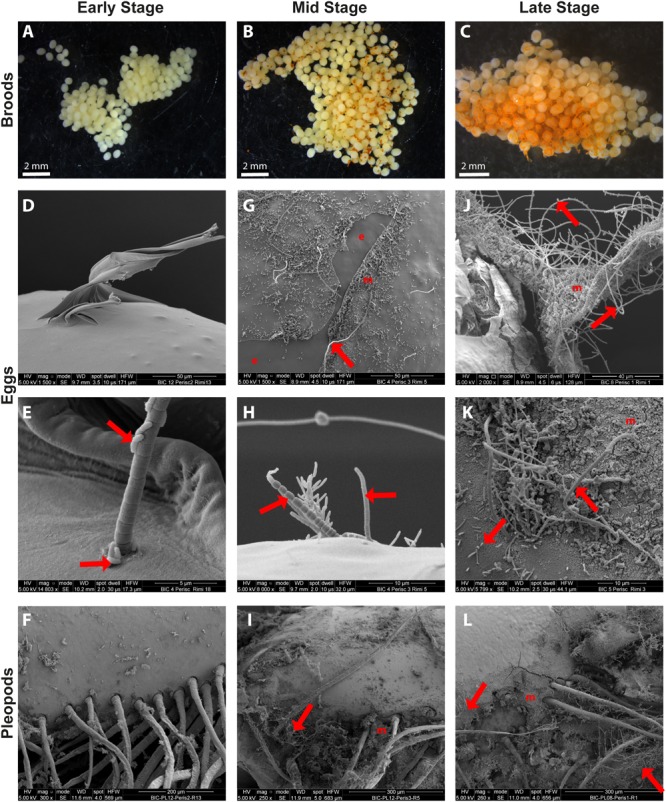
SEM observations of epibiotic bacteria on the surface of R. exoculata eggs and pleopods. Rimicaris exoculata egg broods observed under a stereomicroscope with eggs at (A) early stage, (B) mid stage, (C) and late stage. Individual eggs observed under a scanning electron microscope with eggs at (D,E) early stage, (G,H) mid stage (J,K) and late stage. Pleopods observed under a scanning electron microscope holding broods at (F) early stage, (I) mid stage, (L) and late stage. The crusts observed in the close-ups both on eggs (K) and pleopods (L) in SEM are deposits of ferric oxide, which correspond to the orange coloration observed under stereomicroscope on egg broods (B,C). Red arrows are targeting some of the rod shaped and filamentous bacteria observed in these pictures. e, bare egg envelope areas where mucus layer have been removed and m, egg envelope areas covered by mineral crusts.
