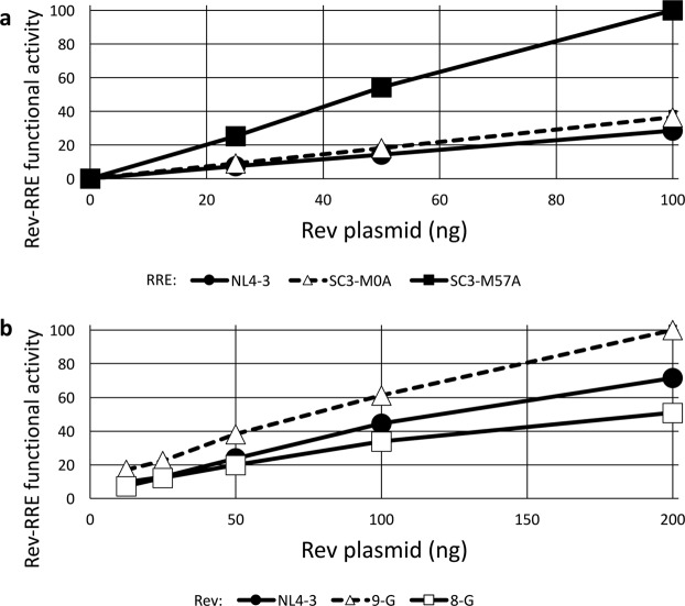
Figure 4.
Differential activity of RRE and Rev sequences. (a) Three different 234-nt RREs were cloned into the eGFP/TagBFP HIV-derived construct, including RREs from NL4-3 and two derived from patient isolates (SC3-M0A and SC3-M57A)23. 1000 ng of the HIV-derived construct was transfected into 8 × 10⁵ 293T/17 cells along with 0, 25, 50, or 100 ng of pCMV-SC3-(M0-B/M57-A) Rev23. Twenty-four hours after transfection, flow cytometry was performed and the ratio of eGFP/TagBFP signal was determined as a measure of Rev-RRE functional activity. The maximum measured activity was set as 100. (b) Three Rev sequences from NL4-3 or two different primary isolates53,54 were cloned into the MSCV vector carrying an mCherry fluorescent marker. 1000 ng of an eGFP/TagBFP HIV-derived construct containing an NL4-3 RRE sequence was transfected into 8 × 10⁵ 293T/17 cells along with 12.5, 25, 50, 100, or 200 ng of a pMSCV-Rev-IRES-mCherry construct. Flow cytometry was performed 24 hours after transfection and the data were analyzed as above. N = 2, bars, which are too small to be seen in this figure, represent SEM for both graphs.

