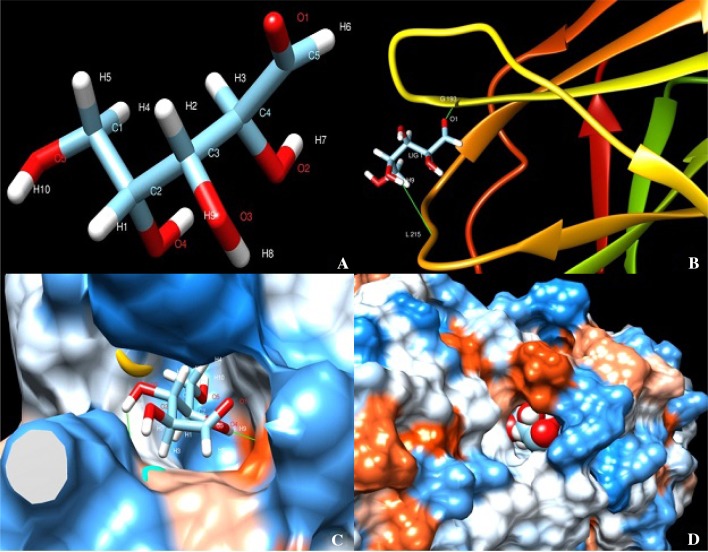
Fig. 3.
Protein–ligand docking using Swissdock viewed in USCF Chimera. a Xylose ligand. b Representation of docking in IK 76-81 using molecular back bone structure. c, d Protein–ligand interaction in NG 77-18 and Co 86032 represented using sphere surface model representation of protein–ligand interaction

