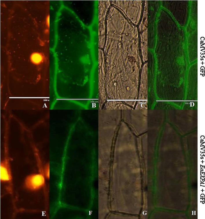
Fig. 5.
Subcellular localization of EaEXPA1 in epidermal onion cells. Both control plasmid (35 s + mGFP5) and EaEXPA1 fused with mGFP5 (35 s + EaEXPA1 + mGFP5) were bombarded in epidermal onion cells. Above figure shows propidium iodide staining (a, e), GFP fluorescence (b, f), bright field (c, g), merged picture with GFP fluorescence and bright field (d, h) in which the expression of the expansin gene in the cell wall can be clearly observed

