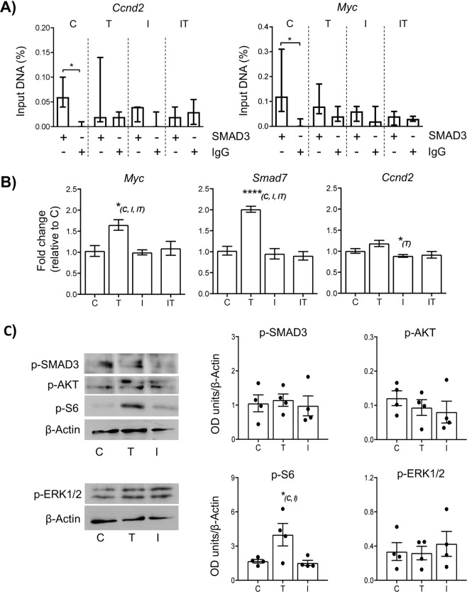
Figure 4.
Short term exposure to TGFβ1 promotes mTOR signalling in neonatal mouse ovaries. Whole neonatal mouse ovaries (d4) were maintained in vitro and exposed for 2 hours in either 1 µM DMSO (control; C), 10 ng/ml of TGFβ1 ligand (T), or 1 µM A83-01 inhibitor (I). An additional group was exposed to 1 µM A83-01 inhibitor followed by 10 ng/ml of TGFβ1 ligand (IT). Ovaries were analysed for gene and protein analysis immediately following treatment. (A) Relative quantification of SMAD3 binding to Ccnd2 and Myc gene promoters in cultured ovaries by ChIP-qPCR. Each group contained 5 ovaries (n = 5). Data are medians ± interquartile range and statistical differences are relative to IgG control (*P < 0.05). (B) Relative expression of Myc, Smad7 and Ccnd2 mRNA in cultured ovaries. Each group contained 5 ovaries (n = 5). Data are means ± SEM; *P < 0.05 vs group indicated in parentheses. (C) Expression of phospho-intermediates in the canonical TGFβ (p-SMAD3), PI3 kinase/Akt (p-AKT), p44/42 MAPK (p-ERK1/2) and mTOR (p-S6) pathways in cultured ovaries by western blot. Example blots are shown (left-hand panels; originals provided in Fig. S4) and levels quantified by normalization against beta-actin (loading control). Data represents four samples from each group, each from an independent blot. *P < 0.05 vs group indicated in parentheses.

