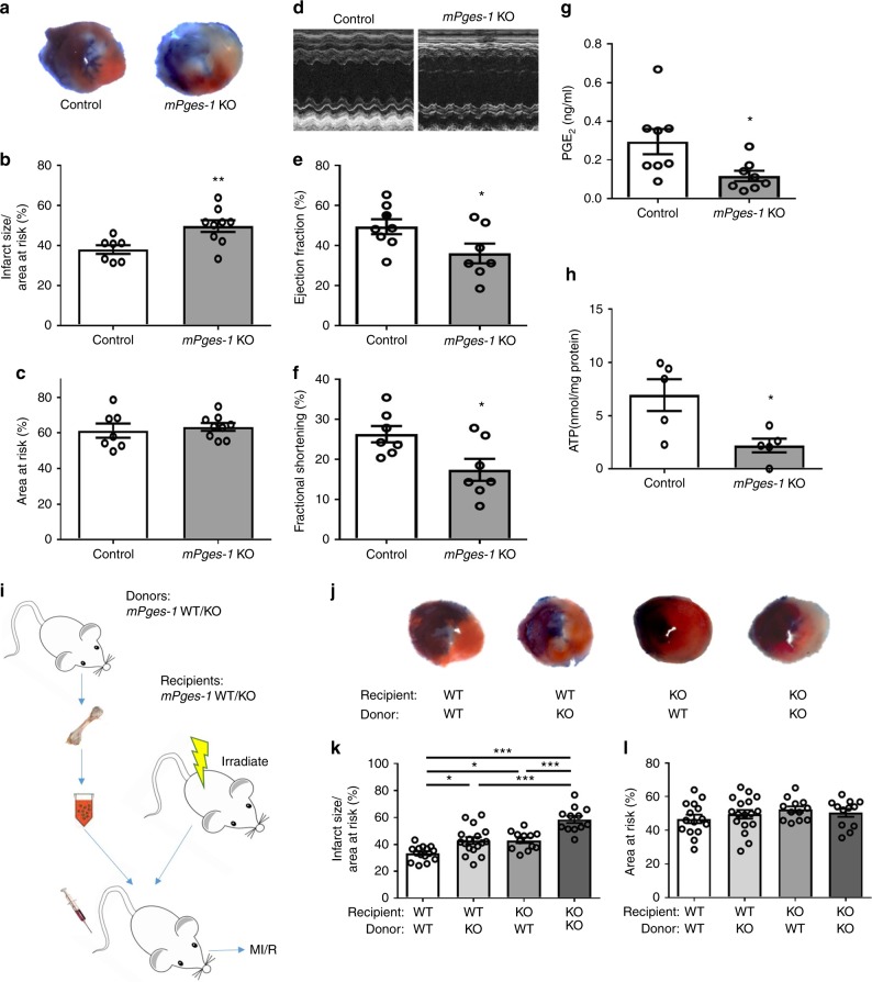
Fig. 2.
Impact of mPges-1 deletion on MI/R injury. Mice were subjected to MI/R injury as detailed in the Methods. a Representative photographs of TTC stained sections from Evans blue perfused hearts. Infarct size (b) and AAR (c) were quantified for mPges-1 KO and control mice. Representative echocardiograph (d), ejection fraction (EF) (e), and fractional shortening (FS) (f) were shown. PGE2 levels were compared (g). Cardiac levels of ATP post MI/R were measured (h). Unpaired Student’s t test (b, c, n = 7, 9; e, n = 8, 7; f, n = 7, 7; g, n = 8, 8; h, n = 5). Bone marrow cells were transplanted reciprocally between mPges-1 WT and KO mice (i). The resulting chimeric mice underwent MI/R injury. Representative TTC staining was shown for each group (j). Infarct size (k) and AAR (l) were analyzed with use of one-way ANOVA and Tukey’s multiple comparison test (n = 15, 17, 12, 12). Error bar indicates SEM

