Figure 4.
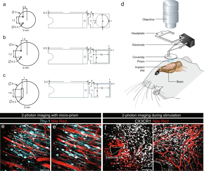
Concomitant 2-photon imaging and brain stimulation. (a–c) Three phases of design of the coverslips and head plates tested during the optimization of the intravital imaging protocol with the selected design illustrated in (c). (d) Schematic of the imaging set-up that was developed to overcome physical challenges such as the size of the objective, cranial window and electrode positioning. (e,f) Representative images of intravital 2-photon imaging in mice expressing CFP under control of the Thy-1 promoter (e-e’) and GFP under the control of the CX3CR1 promotor (f-f’) allowing identification of neurons and microglial cells respectively. (e-e’) Representative images of intravital 2-photon imaging of neurons using microprisms in the absence of stimulation and of (f-f’) microglia 2 weeks after implantation of the electrodes. In both conditions, blood vessels were visualized by injection of Nile red into the orbital vein of mice just prior to imaging. The electrode tip is delineated by a dotted line. Mice were implanted with electrodes 2 weeks prior to the commencement of imaging. Scale bars (e-e) = 50 µm, (f-f’) = 100 µm.

