Fig. 5.
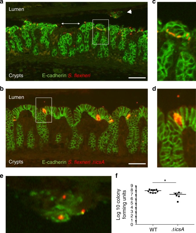
Shigella flexneri spreads from cell to cell in an IcsA-dependent manner. a, b Representative images of colon sections infected with S. flexneri (a) and the ΔicsA mutant (b). Scale bars, 50 μm. E-cadherin, green; S. flexneri, red. c, d Zoom-in on the boxed area in a, b, respectively. e Zoom-in on the area indicated by the arrowhead in a. f Graph showing counts of colony-forming units in the distal colon of animals infected with S. flexneri (wild-type (WT)) or the ΔicsA mutant. Statistical analysis: unpaired t test. ΔicsA vs. S. flexneri (WT), *P < 0.05. Source data are provided as a Source Data file

