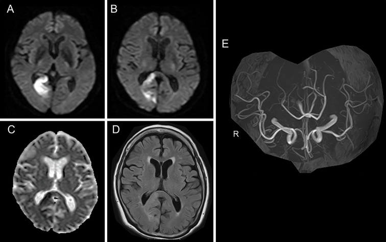
Figure 3.
Magnetic resonance imaging of Case 2. DWI (A, B), ADC mapping (C), FLAIR (D), and MR angiography (E) on Day 2. No lesion was present on the left side of the SCC (B-D). ADC: apparent diffusion coefficient, DWI: diffusion-weighted imaging, FLAIR: fluid-attenuated inversion-recovery, MRA: magnetic resonance angiography, SCC: splenium of the corpus callosum

