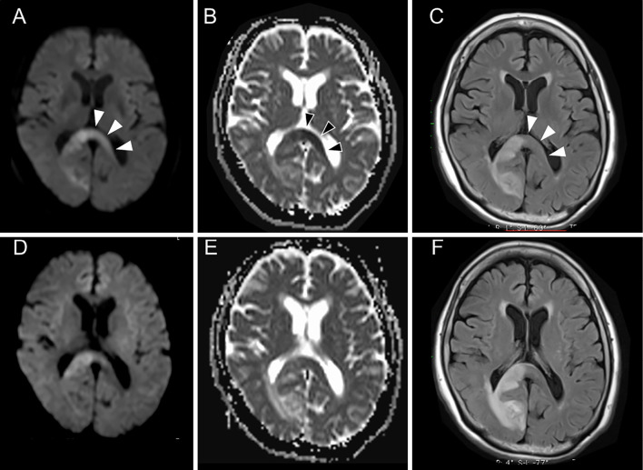
Figure 4.
Follow-up magnetic resonance imaging of Case 2. A hyperintense lesion on DWI (A) and FLAIR (C) with decreased ADC values (B) extended to the left side of the SCC on Day 7 (arrowheads). This lesion on the left side of the SCC became obscure on DWI (D), ADC mapping (E), and FLAIR (F) on day 15. ADC: apparent diffusion coefficient, DWI: diffusion-weighted imaging, FLAIR: fluid-attenuated inversion-recovery, SCC: splenium of the corpus callosum

