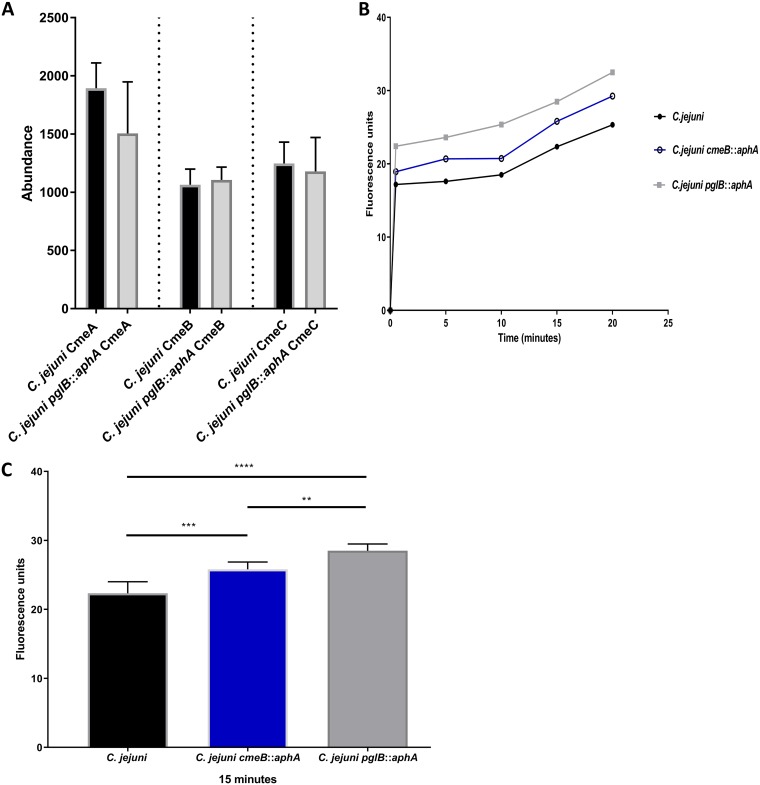
FIG 2.
Differential expression and functional analysis of CmeABC in C. jejuni. (A) Differential expression of CmeABC in wild-type C. jejuni and C. jejuni pglB::aphA. Data are from three biological replicates, and error bars represent standard deviations. Data were analyzed by Student’s t test. (B) Ethidium bromide accumulation test in C. jejuni strains. Thirty milliliters of brucella broth was separately inoculated with overnight cultures of C. jejuni (black circles), C. jejuni cmeB::aphA (open circles), and C. jejuni pglB::aphA (gray squares) to an OD600 of 0.1. Cells were grown until an OD600 of 0.4 to 0.5 was reached, spun down, washed, and resuspended to an OD600 of 0.2 in 10 mM sodium phosphate buffer (pH 7). Cells were then incubated in a VAIN for 15 min at 37°C, and then ethidium bromide was added to a final concentration of 0.2 mg/ml. Fluorescence was read at excitation and emission for 20 min at 37°C. (B) Ethidium bromide accumulation in C. jejuni strains throughout 20 min. (C) Ethidium bromide accumulation in C. jejuni strains at 15 min. The data are means for three biological replicates, two technical replicates each, and the error bars represent standard deviations. Significance was calculated using one-way ANOVA test with multiple comparison and indicated by asterisks as follows: **, P < 0.01; ***, P < 0.001; ****, P < 0.0001.

