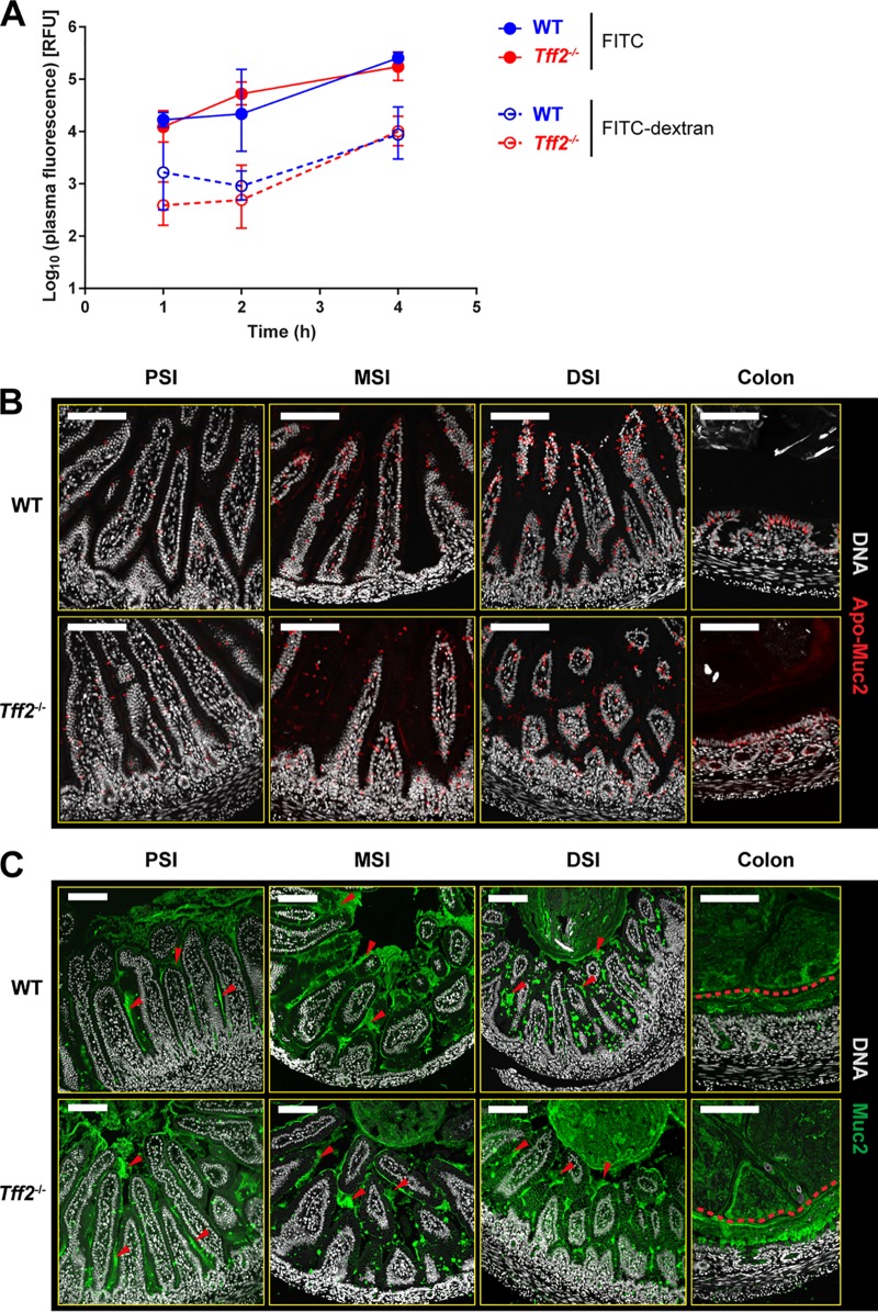
FIG 4.
Loss of Tff2 does not affect GI permeability or the appearance of the GI mucus layer. (A) Intestinal permeability in noncolonized P9 wild-type and tff2−/− pups; plasma fluorescence was determined for 4 h following oral administration of FITC or FITC-dextran. Plasma fluorescence was expressed as log10 relative fluorescence units (RFU). (B and C) Representative confocal micrographs of methacarn-fixed PSI, MSI, DSI, and colonic tissue sections from noncolonized P9 WT and tff2−/− rats obtained 48 h after colonization with E. coli A192PP at P9 and probed for Apo-Muc2 (B) and Muc2 (C). In panel C, the red arrowheads in the small-intestine images show secreted Muc2, and the red dashed lines in the colonic images shows the approximate border of the inner mucus layer. Scale bars, 100 μm.

