
FIG 3.
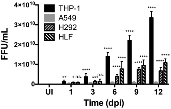
C. burnetii replication differs by cell type. THP-1, A549, and H292 cells and HLFs were infected with C. burnetii for 1, 3, 6, 9, or 12 days. At each time point, samples were harvested, sonicated, and incubated with Vero cells for 4 days; then they were processed for fluorescence microscopy using an antibody against C. burnetii in triplicate in order to quantify the bacterial focus-forming units (FFUs). UI, uninfected cells. *, P < 0.05; **, P < 0.01; ***, P < 0.001; ****, P < 0.0001; n.s., not significant. C. burnetii foci are present in the highest numbers in THP-1 cells, followed by HLFs, H292 cells, and A549 cells, respectively. From day 6 to day 12, THP-1 cells, H292 cells, and HLFs contained significantly higher numbers of C. burnetii than A549 cells, indicating more-efficient replication in nonalveolar epithelial cells.