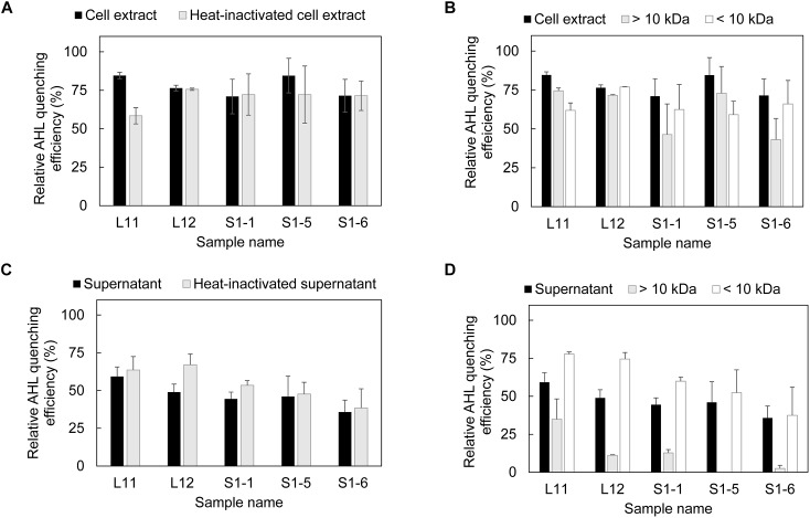
FIGURE 1.
Presence of AHL quenching enzymes in each fraction of AHL quenching bacteria. (A) Evaluation of AHL quenching activity in cell extracts and heat-inactivated cell extracts, (B) separation of cell extract based on molecular size (10 kDa) and evaluation of the AHL quenching effect in each fraction, (C) detection of AHL quenching activity in bacterial supernatant and heat-inactivated supernatant, and (D) separation of bacterial supernatant based on the molecular size and evaluation of the AHL quenching effect in each fraction. Each fraction was mixed with AHL mixture (0.15 mM C4-HSL, 0.13 mM C6-HSL, 0.11 mM C8-HSL and 0.08 mM 3-oxo-C12-HSL) and incubated at 37°C for 18 h for residual AHL determination using a biosensor. The relative AHL quenching efficiency of each sample was calculated based on Eq. (1), as stated in the section “Materials and Methods.” PBS buffer and AS were used to replace the cell extract and supernatant to create the negative control. Three biological replicates were performed, and the results were expressed as the means ± standard error.

