
Figure 3.
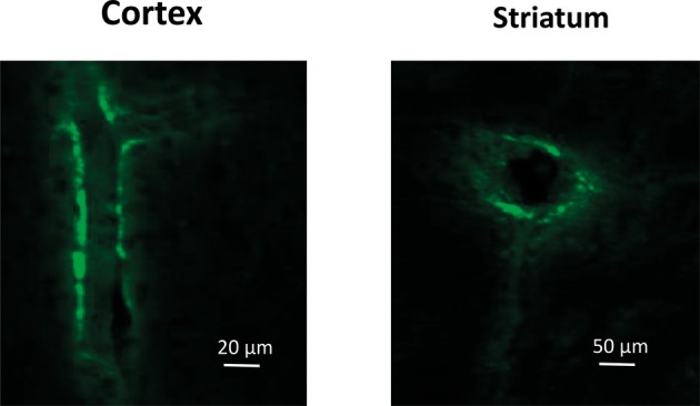
Confocal microscopy images of the blood vessels in the Cortex (lengthwise projection) and Striatum (cross-cut projection) of LPS-treated mice prepared 72 h after intravenous injection of GFP-labeled mMSCs.
Official websites use .gov
A
.gov website belongs to an official
government organization in the United States.
Secure .gov websites use HTTPS
A lock (
) or https:// means you've safely
connected to the .gov website. Share sensitive
information only on official, secure websites.
Confocal microscopy images of the blood vessels in the Cortex (lengthwise projection) and Striatum (cross-cut projection) of LPS-treated mice prepared 72 h after intravenous injection of GFP-labeled mMSCs.