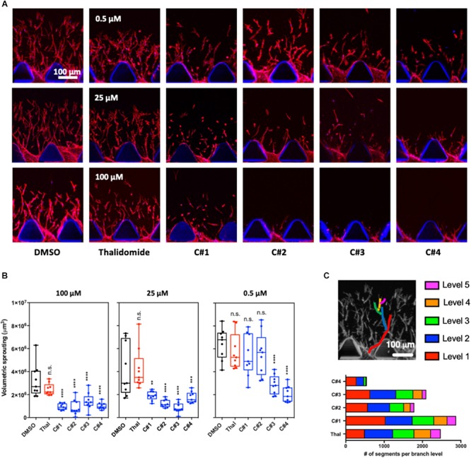
FIGURE 2.

3D angiogenesis assay in microfluidic device. (A) Representative confocal images of the angiogenic sprouting in the 3D extracellular matrix-like hydrogel. Human endothelial cells were treated with Thalidomide or one of the compounds at 0.5, 25, and 100 μM for 72 h. Treatment with vehicle was used as control. Cells are stained for F-actin and Hoechst to visualize actin cytoskeleton filaments and nuclei, respectively. (B) Analysis of the volumetric sprouting for each condition. The box and whiskers plots show all the data points quantified for at least three devices per condition. Statistics were calculated by one-way ANOVA. (C) The different network complexity for each compound or for Thalidomide was analyzed by quantifying the branch levels and the number of segments for each branch level combining all the concentrations for each compound. n.s. = not significant; **p < 0.01; ***p < 0.001; ****p < 0.0001.
