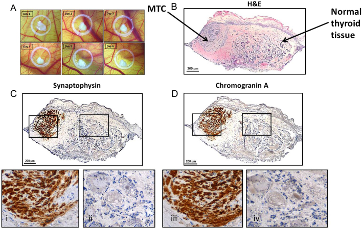
Figure 3.
MTC primary tumour tissue grafted on the CAM retained to express specific tumour markers. (A) Tumour tissue grown on the CAM for 6 days, bar = 1 mm. (B) Subsequent histological analyses showed that the patient sample was composed of MTC and healthy thyroid tissue. The tumour tissue was positive for tumour markers synaptophysin (C and i) and chromogranin A (D and iii), whereas surrounding tissue (ii and iv) showed non-pathological thyroid morphology.

 This work is licensed under a