
Figure 2.
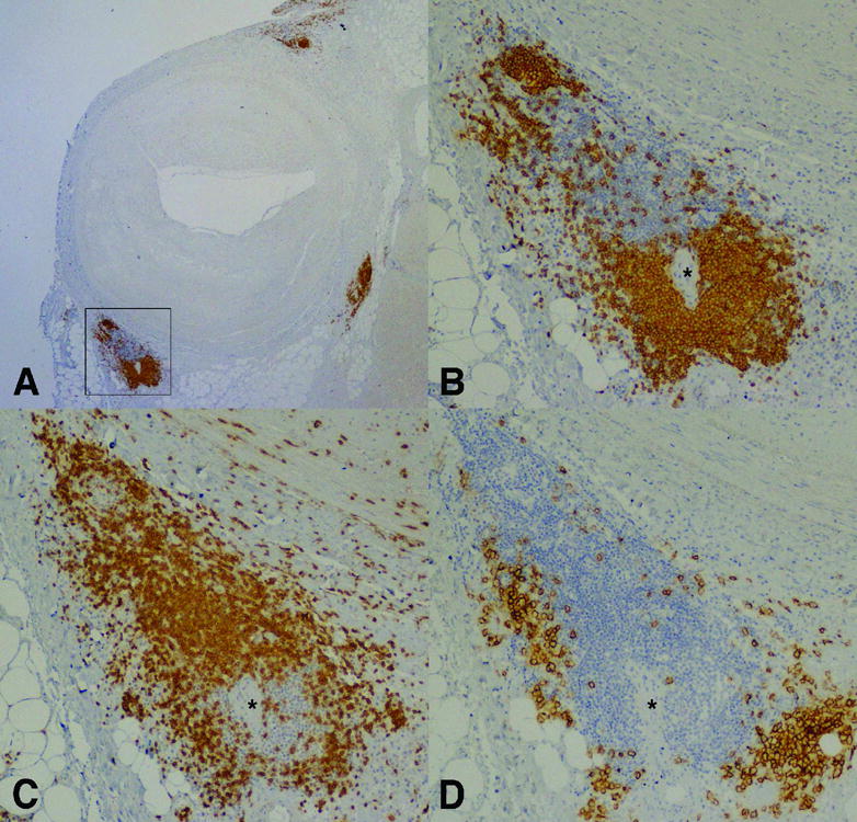
Epicardial coronary artery with AV and adventitial nodular mononuclear cell infiltrates showing compartmentalization of B and T lymphocytes as well as plasma cells. Serial sections are stained by immunoperoxidase for B lymphocytes (CD20; A&B), T lymphocytes (CD3; C) and plasma cells (syndecan-1; D). (Magnification: A; 2X and B–D; 20X) * = arteriole.