
Figure 3.
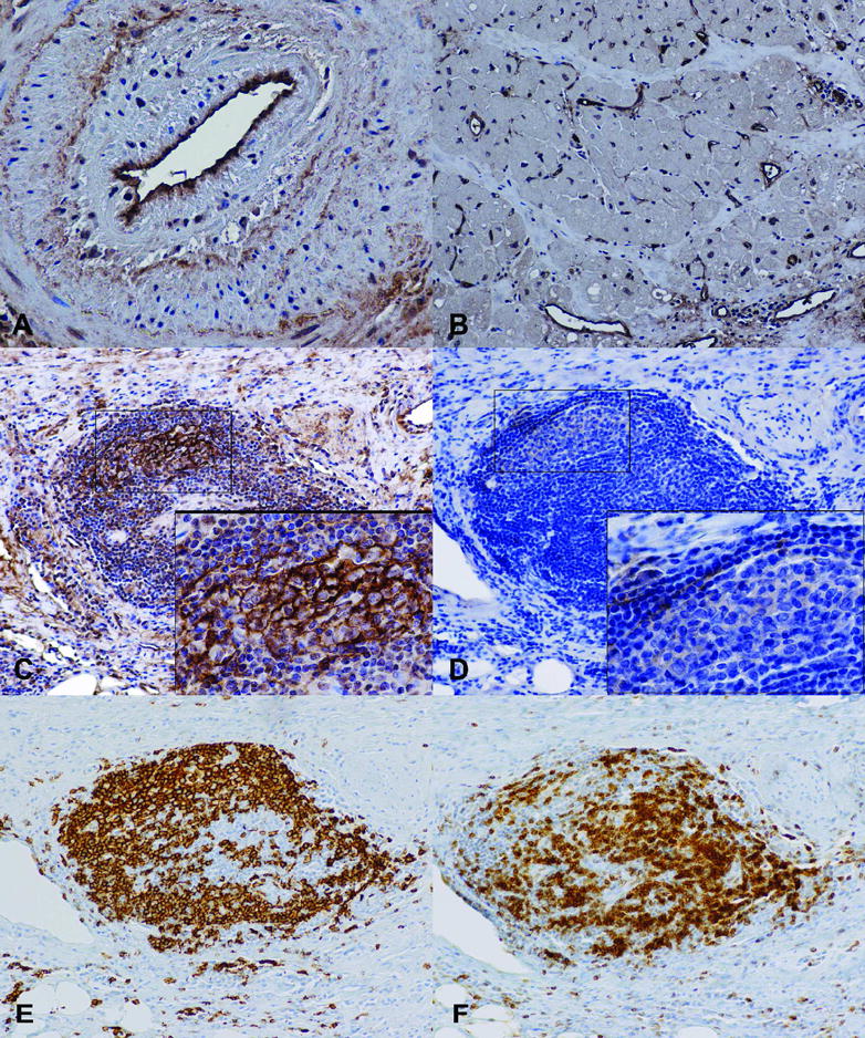
Immunohistochemistry stains of C4d deposition and CXCL13 in patients with AV. C4d deposits are shown on the endothelium of a coronary artery with AV (A) and capillaries of adjacent myocardium (B). Lymphoid nodules stained for C4d (C), CXCL13 (D), B cells (CD20; E), and T cells (CD3; F). (Magnification: A −40X; B–D −20X; Insets − 60X)