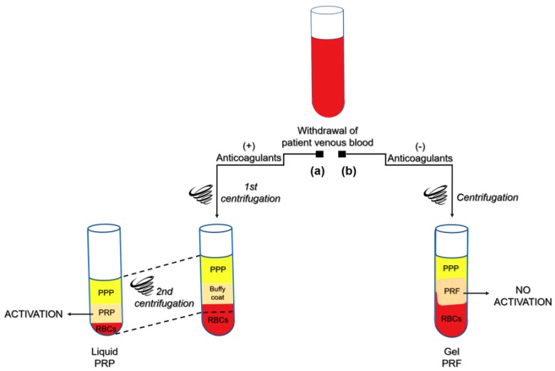
Figure 1.
Platelet concentrates preparation protocols. Schematic drawing of the classical preparation protocols of PRP (Platelet Rich Plasma) and PRF (Platelet Rich Fibrin) hemocomponents. According to PRP protocol (a), blood is collected by venipuncture in the presence of anticoagulants. Thereafter, a two-step centrifugation procedure occurs. The first centrifugation yields three layers: RBCs (red blood cells), “buffy coat” and PPP (platelet poor plasma); hence, PPP and “buffy coat” are transferred into another tube and centrifuged again. After discarding the PPP fraction, the resulting PRP is suspended and activated by fibrin. As regards PRF protocol (b), venous blood is withdrawn without anticoagulants and centrifuged, causing the coagulation and stratification of blood components. In the middle of the tube, between the PPP and the RBC layers, a PRF clot develops, which naturally entraps platelets, leucocytes and molecules like growth factors and fibronectin. To be considered “platelet rich”, hemocomponents should be 5 times concentrated in platelets. Thus, the drawing is intended to be representative of the PRP and PRF manufacture steps, as many variations of the protocols are reported in the literature. There is general consensus in referring to Choukroun’s protocol [3] as the first method for PRF development.

