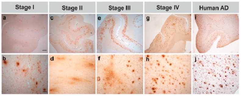
Figure 6.
Photomicrographs exemplifying the four stages of amyloid beta (Aβ1-16) deposition in aged canines’ prefrontal cortex. (a,b) Stage I—very few small plaques with poorly defined borders. (c,d) Stage II—abundant, cloud-like plaques in the deep cortical layers. (e,f) Stage III—abundant, cloud-like plaques in the deep cortical layers and small, distinct plaques in facile layers. (g,h) Stage IV—meager, condensed plaques found in all cortical layers, except layer I. (i,j) Immunoreactivity of amyloid plaques in human’s frontal cortex (patient with Alzheimer’s disease). Bars = 1000 µm in (a,c,e,g,i), and 100 µm in (b,d,f,h,j) [75]. Reprinted from Journal of Alzheimer’s disease, 52, Schütt T., Helboe L., Pedersen L.O., Waldemar G., Mette B., Pedersen J.T.: Dogs with Cognitive Dysfunction as a Spontaneous Model for Early Alzheimer’s Disease: A Translational Study of Neuropathological and Inflammatory Markers, 433–449, 2010, with permission from IOS Press.

