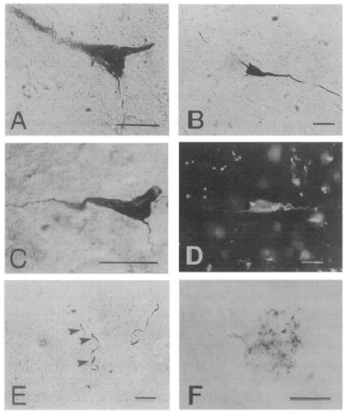
Figure 7.
Neurofibrillary degeneration in the sheep cortex as viewed under the light microscope. Neurofibrillary tangle-like structures (A–D) in sheep brains, stained using: (A) Alz-50 antibody, (B) PHF-I antibody, (C) Gallyas’ silver stain, and (D) thioflavine S. Lesions resembling ‘neuropil threads’ (E) and neuritic plaques (F) could be visualized using PHF-I (shown here) as well as Alz-50. Bars in A = 20 µm, in F = 50 µm. Reprinted from Neuroscience Letters, 170, Nelson P.T., Greenberg S.G., Saper C.B. Neurofibrillary tangles in the cerebral cortex of sheep, 187–190, 1994, with permission from Elsevier [88].

