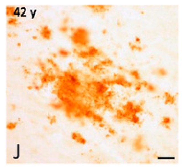
Figure 10.
High-magnification photomicrographs showing a diffuse APP/Aβ-ir (immunoreactive) plaques in the frontal cortex of a 42-year-old female gorilla. Bar = 25 μm. Reprinted from Neurobiology of Aging, 39, Perez S.E., Sherwood C.C., Cranfield M.R., Erwin J.M., Mudakikwa A., Hof P.R., Mufson E.J. Early Alzheimer’s disease-type pathology in the frontal cortex of wild mountain gorillas (Gorilla beringei beringei), 195–201, 2016, with permission from Elsevier [97].

