Figure 6.

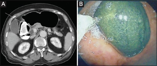
(A) Contrast-enhanced computed tomography showing a duodenal phytobezoar (arrow). (B) Phytobezoar (5×10 cm) fragmentation in the stomach with an ordinary oval 30 mm polypectomy snare (endoscopic view)

(A) Contrast-enhanced computed tomography showing a duodenal phytobezoar (arrow). (B) Phytobezoar (5×10 cm) fragmentation in the stomach with an ordinary oval 30 mm polypectomy snare (endoscopic view)