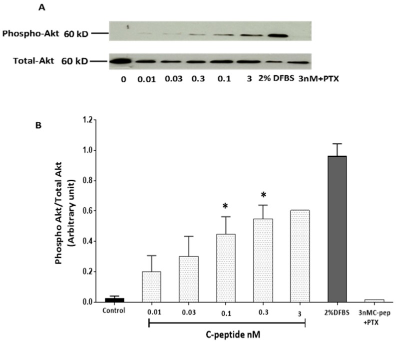
Figure 2.
Dose response study of Akt activation by rat C-peptide in L6 cells. Cells were stimulated with rat C-peptide for 5 min in DMEM. Pertussis toxin (PTX) denotes the effect of co-incubation with 100 ng/mL Pertussis toxin. Phosphorylation of Akt was determined by the Western blot (A) using specific anti-phospho-Akt antibody. DFBS (2%) was used as a positive control. As a loading control, membranes were reprobed with antibody against total Akt; (B) quantification by densitometry of data from three such experiments: mean ± SEM * p < 0.05 versus the unstimulated control.

