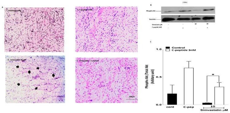
Figure 4.
(A) C-peptide protects against simvastatin-induced cell toxicity. L6 myoblasts were serum starved in DMEM (i); treated with 3 nM C-peptide alone (ii); treated with simvastatin 10 μM (iii); or treated with simvastatin in the presence of 3 nM rat C-peptide (iv) for 72 h. Morphological visualization was assessed by using the Wright stain and light microscopy. Black arrows indicate shrunken cells (consistent with apoptosis). Magnification ×200; (B) C-peptide blunts the inhibitory effect of simvastatin on phospho-Akt activation in L6 myoblasts. Cells were treated with 10 µM simvastatin and co-incubated with 3 nM rat C-peptide for 72 h. Phosphorylation of Akt was determined by the Western blot using specific anti-phospho Akt antibody. As a loading control, membranes were reprobed with antibody against total Akt (shown in the lower blot); (C) densitometric analysis of four experiments. Data are presented as mean ± SEM * p < 0.05.

