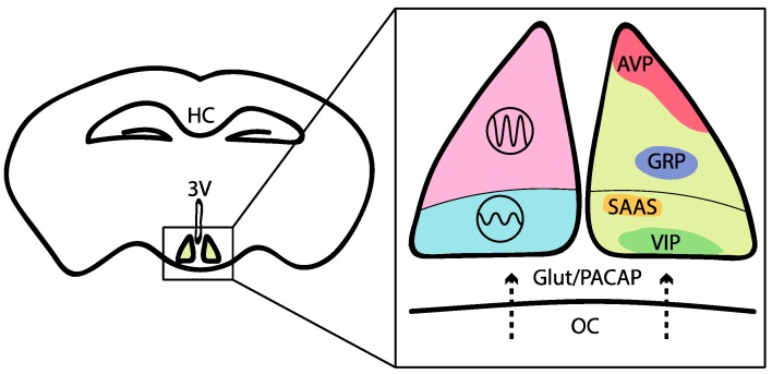
Figure 2.
Anatomy of the suprachiasmatic nucleus. A schematic of a coronal slice of the mouse brain showing the location of the suprachiasmatic nucleus (SCN) in the hypothalamus above the optical chiasma (OC). The detailed portion shows the distinct anatomical sections of the SCN. The dorsal and ventral SCN are in pink and blue respectively. Subsets of SCN neurons are shown on the right: GABA+ (yellow), VIP+ (green), AVP+ (red), SAAS+ (gold) and GRP+ (purple) are expressed in the dorsal SCN. DRD1a+ (most SCN cells) and NMS+ neurons (~40%) are not shown. HC- Hippocampus, 3V- third ventricle.

