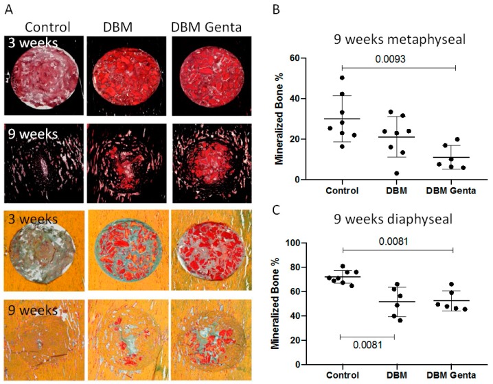
Figure 3.
Exemplary histological pictures of the diaphyseal drill hole defects. Tissue was stained with Safranin O/van Kossa (A, top) to visualize the mineralized bone, or with Movat Pentachrome (A, bottom) to differentiate the tissues. Healing progressed from week three to week nine in all groups, with less healing in the gentamicin-treated defects. (B,C) Histomorphometric analysis: After nine weeks, significantly less mineralized tissue was formed in the gentamicin-loaded DBM group, both metaphyseal and diaphyseal. Diaphyseally, significantly less bone was formed in the DBM group compared to control. Kruskal-Wallis test with Dunn´s (n = 6–8 per group and time point).

