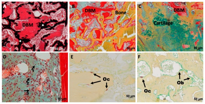
Figure 4.
Safranin O/van Kossa (A), Movat Pentachtome stain (B–D) or TRAP stain (E,F) of drill hole defects after three (A,B,D,E) or nine (C,F) weeks healing. (A) Remineralization (black, *) of the implanted DBM (intense red stained tissue with empty lacunae) occurred after three weeks. (B) Active remodeling of the DBM was visible: DBM was attached to and integrated into newly formed bone trabeculae. (C) Endochondral bone formation was only seen in a few drill holes filled with DBM. (D) No difference regarding the abundance of inflammatory cells was detectable between the groups; lymphocytes (arrow 1), macrophages (arrow 2) or histiocytes (arrow 3) were detectable in all groups. (E) Osteoclasts (Oc) were detectable on the surface of the DBM, but also inside DBM after three weeks. F) Shift to the coverage with mostly osteoblasts (Ob) compared to osteoclasts (Oc) at the late time point. Size of scale bars is given in the figures.

