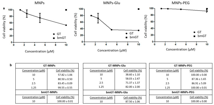
Figure 3.
Biocompatibility of different functionalized nanoparticles with L929 cells. L929 cells were incubated with MNPs functionalized with GT, GT and glucose, and GT and PEG, respectively at different concentrations (1.25, 2.5, 5 and 10 µM) overnight at 37 °C. Subsequently, cells were washed twice with PBS to remove non-internalized MNPs. L929 incubated with the maximum concentration of MNPs functionalized with bmGT, in the presence or absence of glucose or PEG, were used as control. (a) Quantification of cell viability was carried out by MTT as indicated in experimental procedures. The IC50 values were determined by extrapolation, which were presented as mean ± SEM of three independent experiments. Statistical analysis between MNPs functionalized with GT and GT/glucose was performed using paired t test. *, 0.01 < p < 0.05. (b) Analysis of the effect of GT-MNPs, glucose-conjugated GT-MNPs and PEG-conjugated GT-MNPs on the cell viability of L929 cells by Trypan Blue by microscopy. Data are expressed as the mean values ± SEM of three separate experiments.

