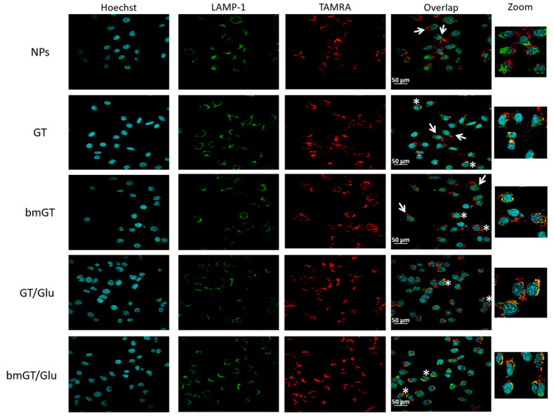
Figure 5.
Localization of MNPs by confocal microscopy. L929 cells were incubated with MNPs or MNPs (10 µM) functionalized with GT, bmGT, GT and glucose, or bmGT and glucose for 4 h at 37 °C. Subsequently, cells were washed and fixed with PFA4% and stained with Hoechst 3342 (DNA marker) and anti-Lamp1 (lysosome marker) followed by a secondary Alexa 488 labelled antibody as indicated in materials and methods. Fluorescence images were taken on a confocal microscope. The overlap shown MNPs internalized (white asterisks) and non-internalized MNPs (white arrows). Higher magnifications (zoom) are shown. Scale bar = 50 μm.

