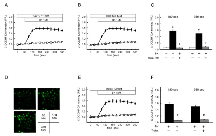
Figure 2.
Bradykinin induces intracellular reactive oxygen species. (A,B) Time course of changes in 6-carboxy-2′ 7′-dichlorodihydrofluorescein di-(acetate, di-acetoxymethyl ester) (C-DCDHF-DA) intensity. Porcine aortic endothelial cells were incubated with modified Tyrode’s solution (control, [Ca2+]o = 1mM, open square; n = 53), and then BK was applied (closed circle; n = 111). HOE 140 (open triangle; n = 107) significantly suppressed the BK-induced increase of C-DCDHF-DA intensity. (C) Summarized data of C-DCDHF-DA intensity at 180 s and 360 s with or without HOE 140. (D) Two-dimensional images of C-DCDHF-DA 60 s (before BK application) (upper left), 180 s (upper right), and 360 s 6 (lower left). (E,F) The same experiments as (A) conducted in the presence (open triangle; n = 185) or absence (closed circle; n = 111) of trolox. Increased C-DCDHF-DA intensity reflects cytosolic reactive oxygen species production. Data are expressed as the mean ± SEM of three independent experiments in separate cell culture wells. * p < 0.01 versus control, † p < 0.01 versus BK, and # p < 0.05 versus BK. Two-factor factorial ANOVA was used for analysis of difference.

