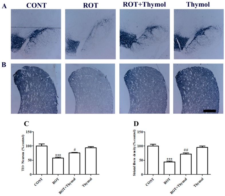
Figure 2.
The illustrative photomicrograph showing expression of TH+ neurons in substantia nigra par compacta (SNc) (A) and TH-ir dopaminergic fibers in striatum (B). The scale bar is 100 µm. The expression of TH+ neurons and TH-ir fibers were reduced in the SNc region of rotenone (ROT) challenged rats as compared to vehicle injected rats in the CONT group. Thymol treatment to ROT challenged rats showed remarkable expressions of TH+ neurons and TH-ir fibers as compared to ROT injected rats. Quantification data showed significant (*** p < 0.001) decrease in the number of TH+ neurons and density of TH-ir fibers in ROT group rats compared to control rats. While thymol treatment to ROT injected rats showed significant (# p < 0.05; ## p < 0.01) increase in TH+ neurons and TH fibers density as compared to ROT alone injected rats (C,D).

