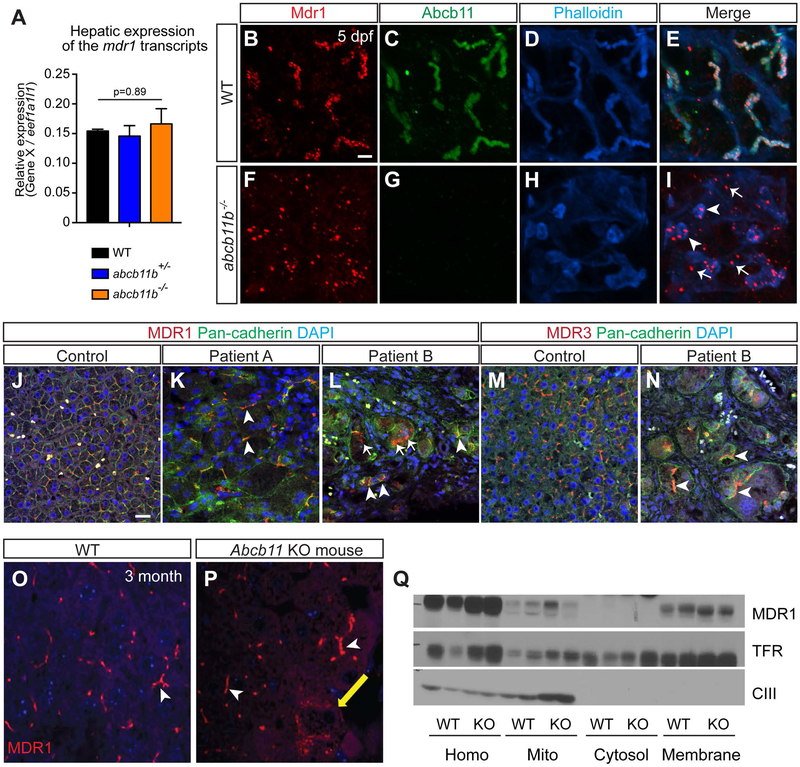
FIG. 5. MDR1 protein was mislocalized in abcb11b mutant zebrafish and a patient with nonsense mutations in ABCB11.
(A) qPCR analysis comparing mdr1 transcript expression in the livers of WT, heterozygous, and homozygous mutant larvae at 5 dpf. Triplicates were performed. The results are represented as relative expression levels that were normalized to the house keeping gene eef1a1l1 (mean±s.e.m.). Statistical significance was calculated by one-way ANOVA and Tukey’s post-hoc test. (B-I) Confocal three-dimensional projections of WT (n=10) and abcb11b mutant (n=10) livers at 5 dpf. (B,F) Mdr1 protein expression; (C,G) Abcb11 protein expression. (D,H) phalloidin staining; (E,I) merged images. Ventral views, anterior to the top. (J-L) Confocal single-plane images of liver sections from control and two PFIC2 patients that were stained with MDR1 antibody (red), Pan-cadherin antibody staining cell membranes (green), and DAPI (blue). Arrowheads in (I,K,L) mark MDR1 expression at the membrane. Arrows in (I,L) mark MDR1 expression in the hepatocyte cytoplasm. (M,N) Confocal single-plane images of liver sections from control and patient B stained with MDR3 antibody (red), Pan-cadherin antibody (green), and DAPI (blue). Arrowheads show that MDR3 was localized at the hepatocyte cell membrane. (B-N) Scale bars, 20 μm. (O,P) Confocal images showing MDR1 protein expression in the livers of WT and Abcb11 knockout mice at 3 month of age. White arrowheads mark MDR1 expression in the canaliculi and yellow arrow marks the expression in the cytoplasm. (Q) Subcellular fractionation assay showing that MDR1 protein is mainly expressed at the cell membrane in both WT and Abcb11 knockout mice. TfR is the transferrin receptor that marks the plasma membrane and CIII represents mitochondrial complex.

