Figure 4.
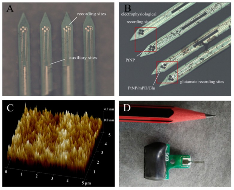
Structure and modification of microelectrode array (MEA). (A) Silicon-based MEA with four shafts [92]. The 16 round sites were used as recording sites to detect electrophysiological and electrochemical signals, and the three rectangular sites were used as auxiliary sites in the three-electrode system. (B) The electrophysiological recording sites are modified with PtNP and glutamate recording sites have three different layers (PtNP/mPD/Gluox) modification. The thicknesses of PtNP layer and the enzyme layer were 3.2 and 1.5 μm, respectively. (C) The AFM photograph of the surface of Gluox enzyme layer. Surface irregularities make Gluox more accessible to glutamate. (D) The physical view after package, the size of this sensor is close to the pencil head and the weigh is about 1 g [68]. (Reproduced here with copyright permission [68]).

