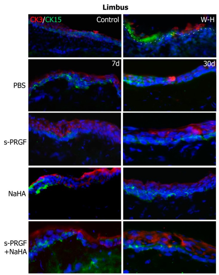
Figure 7.
Fluorescent immunostaining for CK3 (red) and CK15 (green) on the limbal area of rabbit corneas after healing of the epithelial defect. Corneas were treated with s-PRGF, s-PRGF and NaHA, NaHA, or PBS (as a control) and were processed 7 and 30 days after cornea surgery. Control corresponds to a healthy rabbit cornea with no surgery. The W-H image shows a cornea processed 48 h after surgery without complete re-epithelialization. The dotted line shows the limit between the epithelium and stroma layers. Magnification 200×.

