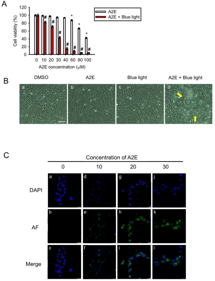
Figure 1.
Blue light damages A2E-laden retinal pigment epithelial (RPE) cells. (A) Cell viability (%) was measured using the 3-(4,5-dimethyl-2-thiazolyl)-2,5- diphenyl-2H-tetrazolium bromide (MTT) assay. A2E-laden RPE cells with or without blue light-emitting diode (LED) light (BLL) treatments were compared with control cells in the dark; 6 independent experiments were performed with three each. White bars indicate RPE cells exposed to 0, 10, 20, 30, 40, 60, 80, and 100 μM synthetic A2E for 12 h. Red bars indicate RPE cells co-treated with BLL for 12 h (460 nm, 150 lux) and the indicated concentration of synthetic A2E (0, 10, 20, 30, 40, 60, 80, and 100 μM). Data represent the average percent cell viability ± SE. * p < 0.05 was considered statistically significant compared to controls. # p < 0.05 compared with the A2E group. (B) Morphology of A2E-laden RPE cells with or without BLL exposure is shown. (a) A representative figure of RPE cells grown to confluence and loaded with dimethyl sulfoxide (DMSO)as a vehicle control. (b) A representative figure of RPE cells grown to confluence and loaded with 30 μM A2E. (c) A representative figure of RPE cells exposed to BLL for 12 h. (d) A representative image of RPE cells co-treated with 30 μM A2E and exposed to BLL for 12 h is shown. RPE cells were observed to have a slim and shrinking morphology (indicated by yellow arrows). Results from 3 independent experiments are shown (N = 3). Scale bar, 100 μm. (C) Photographs showing A2E-laden RPE cell autofluorescence are presented. RPE cells were loaded with the indicated concentration of A2E (10, 20, and 30 μM) for 12 h. Immunofluorescence showing 4′-6-diamidino-2-phenylindole (DAPI) staining (blue) in the cell nuclei and localized autofluorescence (green) in A2E cells is presented. Results from 5 independent experiments are shown (N = 5). Scale bar, 25 μm.

